Cell Biology, Cell Structure
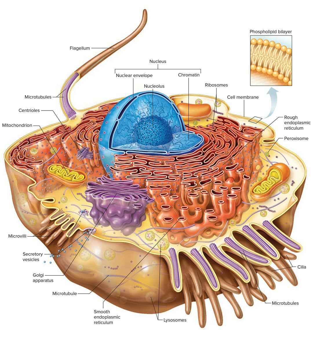
A diagram of a plasma membrane shows a phospholipid bilayer with 3 proteins embedded in the bilayer. One of the proteins is shown with a channel in it. The 3 proteins have lines with the label integral membrane proteins. On the inner side of the phospholipid bilayer is another protein that is positioned up against the inner portion of the bilayer.
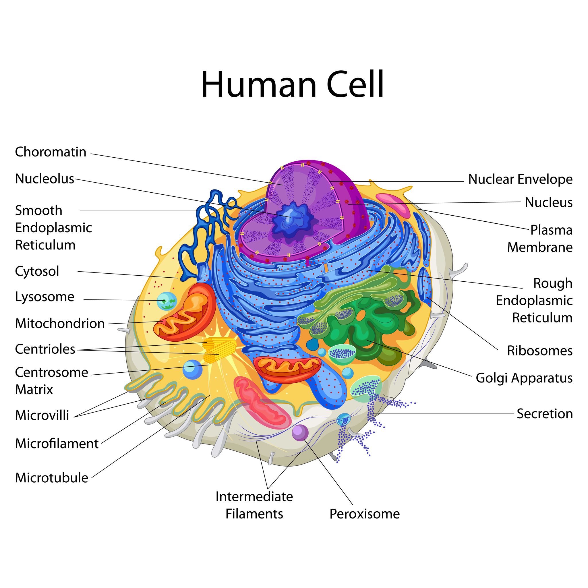
Education Chart of Biology for Human Cell Diagram Best Acupuncture llc
cell, in biology, the basic membrane-bound unit that contains the fundamental molecules of life and of which all living things are composed.A single cell is often a complete organism in itself, such as a bacterium or yeast.Other cells acquire specialized functions as they mature. These cells cooperate with other specialized cells and become the building blocks of large multicellular organisms.
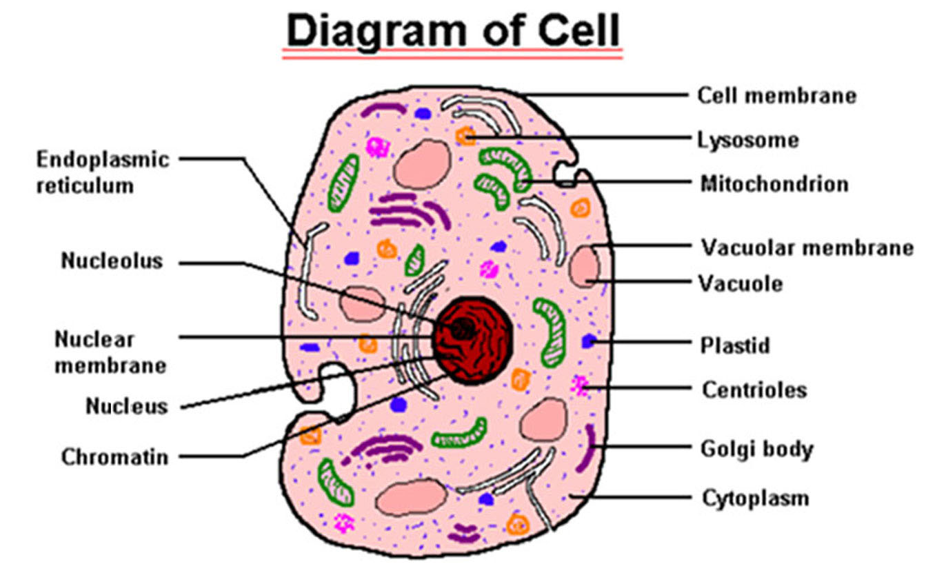
Structure of cell Cell structure and functions, Class 8
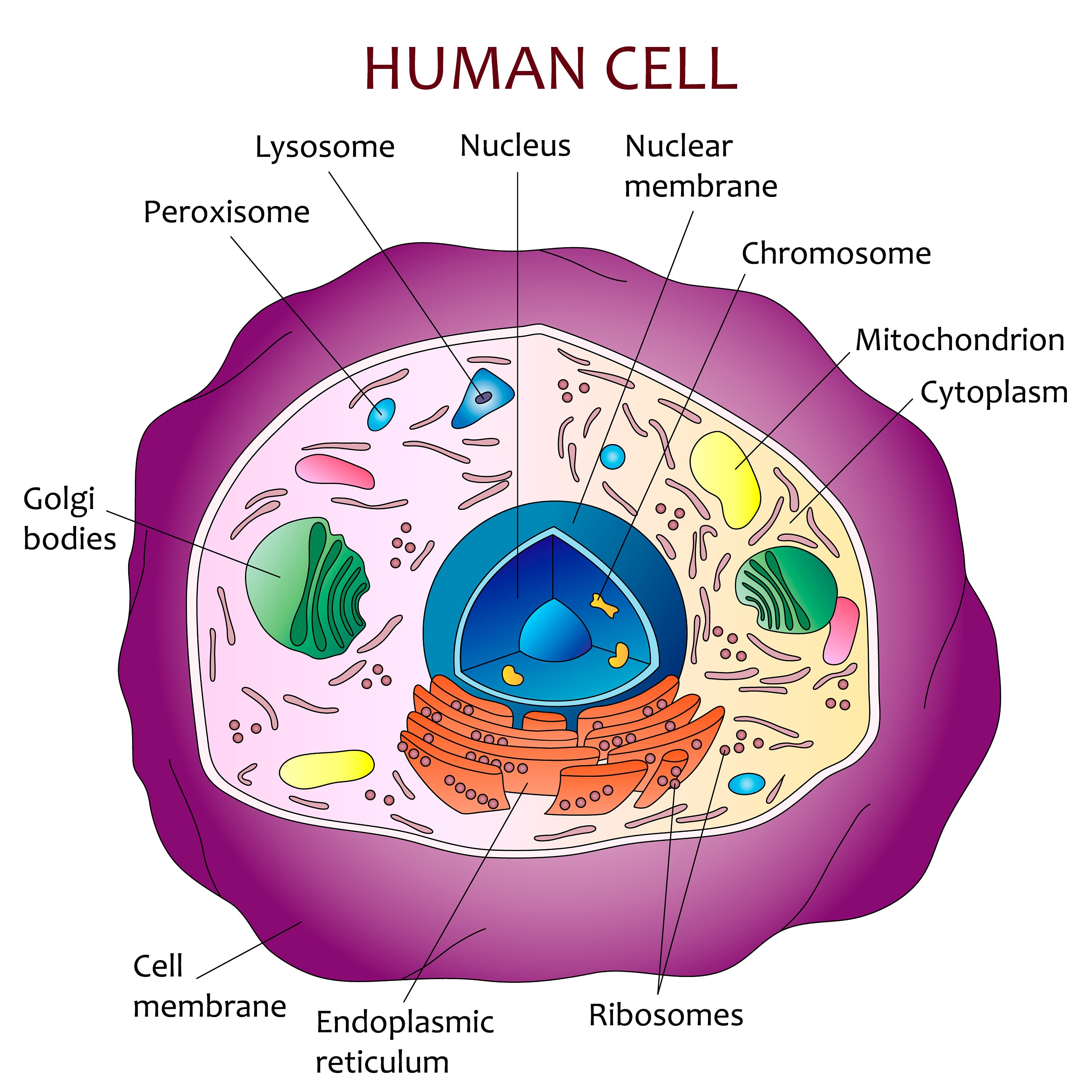
Diagram of the human cell illustrating the different parts of the cell. Cell Membrane The cell membrane is the outer coating of the cell and contains the cytoplasm, substances within it and the organelle. It is a double-layered membrane composed of proteins and lipids.
Education 645 High School Biology
The nucleus is a large organelle that contains the cell's genetic information. Most cells have only one nucleus, but some have more than one, and others—like mature red blood cells—don't have one at all. Within the nucleus is a spherical body known as the nucleolus, which contains clusters of protein, DNA, and RNA.
Human cell diagram Etsy
Cell Membrane The cell membrane is the outer coating of the cell and contains the cytoplasm, substances within it and the organelle. It is a double-layered membrane composed of proteins and lipids. The lipid molecules on the outer and inner part (lipid bilayer) allow it to selectively transport substances in and out of the cell.
Human Cell Diagram 6406474 Vector Art at Vecteezy
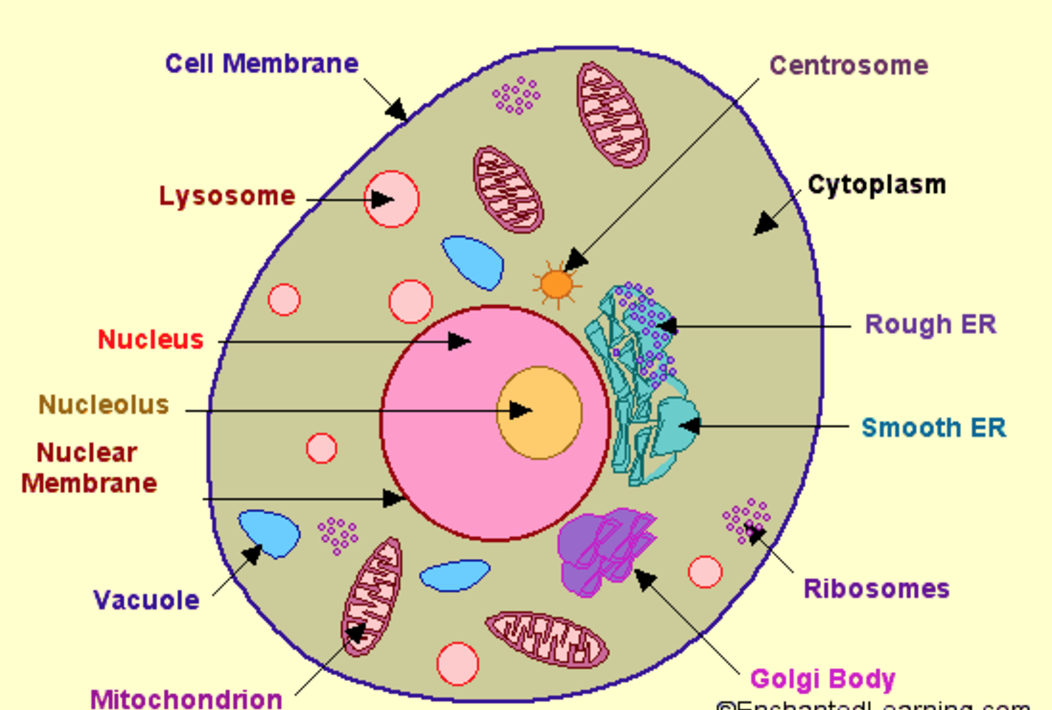
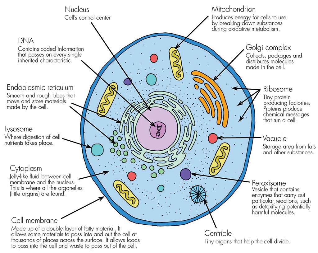
A cell consists of three parts: the cell membrane, the nucleus, and, between the two, the cytoplasm. Within the cytoplasm lie intricate arrangements of fine fibers and hundreds or even thousands of miniscule but distinct structures called organelles. Cell membrane Every cell in the body is enclosed by a cell ( Plasma) membrane.

Pin on Micro
Download the Human Cell Diagram 6406474 royalty-free Vector from Vecteezy for your project and explore over a million other vectors, icons and clipart graphics!

Human Cell Diagram, Parts, Pictures, Structure and Functions
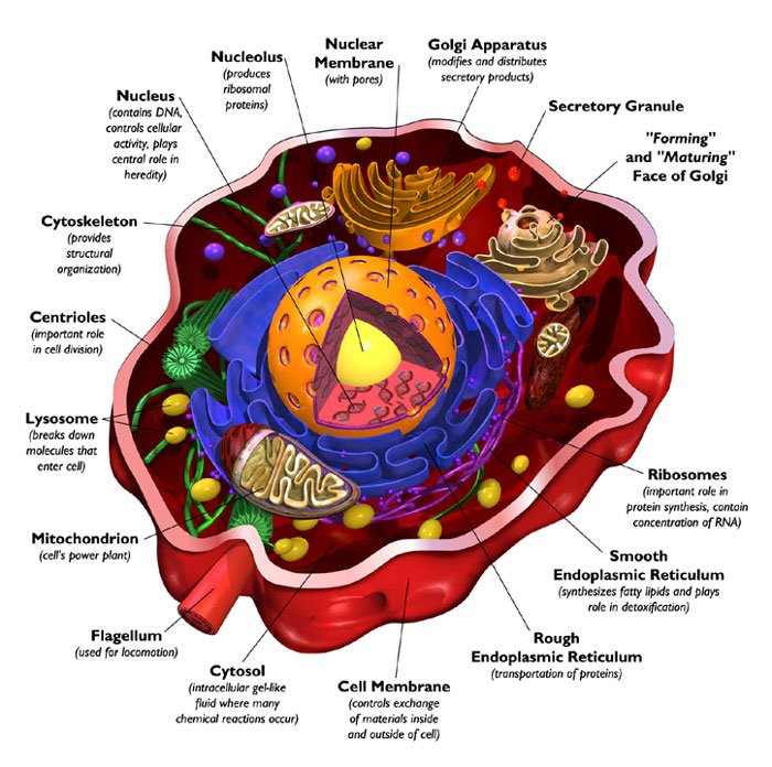
A cell is the smallest living thing in the human organism, and all living structures in the human body are made of cells. There are hundreds of different types of cells in the human body, which vary in shape (e.g. round, flat, long and thin, short and thick) and size (e.g. small granule cells of the cerebellum in the brain (4 micrometers), up to the huge oocytes (eggs) produced in the female.

human cell Diagram Quizlet
During exercise when muscles do not get enough oxygen, lactate is produced. After exercise oxygen dept must be repaid. Slow-twitch muscles rely on aerobic respiration. Fast-twitch fibres generate.

Explain the nucleus of a cell with a neat labeled diagram Science
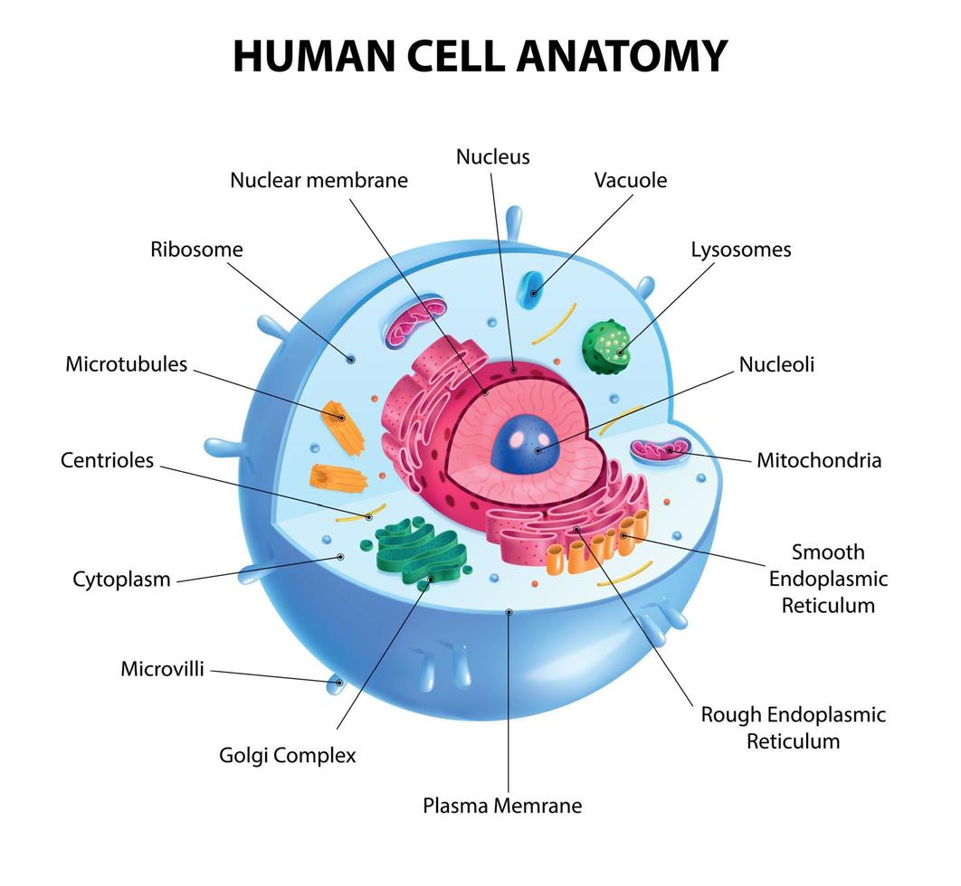
73,930 human cell structure stock photos, 3D objects, vectors, and illustrations are available royalty-free. See human cell structure stock video clips Filters All images Photos Vectors Illustrations 3D Objects Sort by Popular Human cell anatomy infographics with realistic educational chart and labelled parts on white background vector illustration

Cell Structure
The cell is the fundamental organizational unit of life. All living things are composed of cells, which then further subdivide based on the presence or absence of the nucleus, into two types: eukaryotic cells (Greek, Eu=true, karyo=nut, nucleus) - these cells are present in all the human, animal and plants with a clear, distinct nucleus. Prokaryotic cells are some bacteria and blue-green algae.

34 Human Cell Diagram To Label Labels For Your Ideas
Using a cell diagram as the reference point, these quizzes challenge you to label the cell according to the different parts you have just learned about. Cell diagram labeled

Pin on Animal cell
This diagram depicts Human Cell. Human anatomy diagrams show internal organs, cells, systems, conditions, symptoms and sickness information and/or tips for healthy living. This body anatomy diagram is great for learning about human health, is best for medical students, kids and general education.
Cells Haleo
(a) The ER is a winding network of thin membranous sacs found in close association with the cell nucleus. The smooth and rough endoplasmic reticula are very different in appearance and function (source: mouse tissue). (b) Rough ER is studded with numerous ribosomes, which are sites of protein synthesis (source: mouse tissue). EM × 110,000.

identify and label each part of the eukaryotic cell
The cell is the basic unit of any living organ and it is the organ that replicate on its own determining growth. The cell does not need any other triggering element for its multiplication since it is self contained. Cell was first discovered by Robert Hooke in the year 1665. A person is made of […]
Structure Of Human Cell With Labels Images & Pictures Becuo
Key points: All cells have a cell membrane that separates the inside and the outside of the cell, and controls what goes in and comes out. The cell membrane surrounds a cell's cytoplasm, which is a jelly-like substance containing the cell's parts. Cells contain parts called organelles. Each organelle carries out a specific function in the cell.